
Automated breast ultrasound is a secondary screening examination wherein your breasts are examined by health experts. These are most recommended for women with dense breast tissues. If you have signs of breast abnormalities or cancer, you must undergo this procedure. Let’s read about automated breast ultrasound in detail. Click to find out more!
An overview of automated breast ultrasound
Automated breast ultrasound is a medical imaging technique used for breast cancer screening and detection. It produces three dimensional images using ultrasonic waves. In this process, there is no ionizing radiation like mammography does. Thus, it is a safe screening tool beneficial for women with dense breast tissue. Additionally, it is great for those who are at high risk of breast cancer.
Customized ultrasound equipment with an automated scanning device moves around the breast. Further, it takes pictures from various angles. Radiologists can then examine these pictures to find and assess anomalies like lumps, cysts, or other indications of breast cancer. When combined with mammography, ABUS is frequently utilized as an additional screening method to increase the identification of breast cancer, especially in women with thick breast tissue.
What happens before the automated breast ultrasound?
- It is crucial that the imaging specialist review your current study with access to any prior mammograms for comparison. On the day of your exam, kindly bring any prior mammograms.
- Please bring any orders you may have received from your doctor.
- If you have a pacemaker or have been advised to have a follow-up, short-term mammography or ultrasound for breast imaging, this study is not suggested for you.
- Kindly keep your valuables and jewelry at home.
What happens during the automated breast ultrasound?
Here is the step by step process of what happens during the automated breast ultrasound:
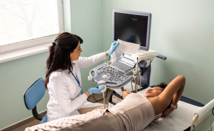
- On the test table, you will be requested to lie on your back.
- Your breast will be rubbed with the lotion. The lotion facilitates the sound waves entering your breast from the machine.
- You will have the cushioned ABUS transducer positioned on your breast.
- Sound waves will reverberate among your breast’s various tissues. This wave action will produce “echoes.” The transducer receives the echoes and reflects them, converting them into electronic impulses.
The impulses are subsequently converted into images by a computer and displayed on a TV monitor. You can watch these moving pictures immediately or record them for later analysis.
- It will take about one hour to complete your exam.
Wrapping up
Automated breast ultrasound has gained popularity nowadays. In this article, we discussed automated-based ultrasound in detail.


